
Aggressive bone lesion in a dog
Case information
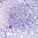
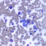
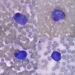
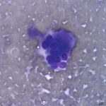
A 7 year old male castrated English Bulldog was presented to Cornell University Veterinary Specialists (CUVS) for further evaluation of an aggressive bone lesion. The dog initially presented to the referring veterinarian for a one week history of lameness. The dog was noted to be painful on palpation of the left elbow and shoulder. Radiographs of the left limb revealed an aggressive lesion in the proximal half of the left humeral diaphysis, distal to the metaphysis, characterized by indistinct / mottled intramedullary and cortical lysis with adjacent periosteal new bone formation on the caudal cortex. There was no apparent soft tissue swelling. Three-view thoracic radiographs were performed, and there was no evidence of metastatic disease. At CUVS, a fine needle aspirate was performed of the bone lesion and submitted for cytologic evaluation.
Evaluate the provided cytologic images and answer the following questions:
1. What are you differential diagnoses for the bone lesion based on the radiographic description?
2. Which of these differentials is favored given your cytologic interpretation?
3. What additional testing is recommended?
|
|
|
|
|
Answer on next page