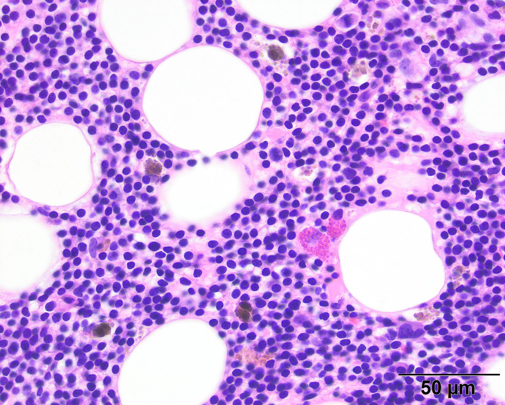
Figure 7: Bone core biopsy, 50x H&E stain
Bone core biopsy, 50x H&E stain. Similar to the aspirate, the marrow is effaced by small tumor cells, with interspersed hemosiderin-laden macrophages and low numbers of hematopoietic precursors (eosinophils are evident in this image). In the biopsy, the plasmacytoid features of the cytoplasm of the cells was not discernible and they resembled small lymphocytes, leading to a histologic diagnosis of chronic lymphoproliferative disease….
Figure 7: Bone core biopsy, 50x H&E stain Read More »