
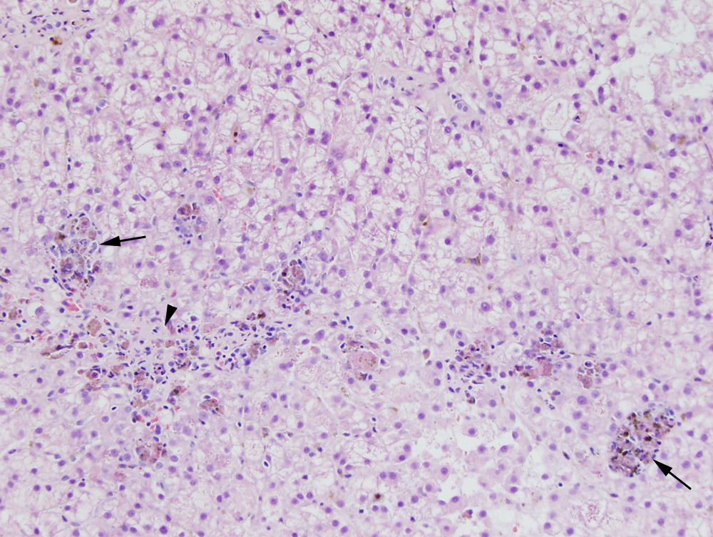
Several aggregates of copper-laden macrophages (copper granulomas) are seen throughout the liver (arrows). Macrophages within the granulomas also contained iron (confirmed with a Prussian blue stain [not shown]) and lipofuscin. The granulomas were mostly found around the central vein (zone 3, arrowhead), along with increased numbers of neutrophils. A small to moderate amount of copper is also seen in the cytoplasm of nearby hepatocytes. The hepatocytes showed diffuse cytoplasmic vacuolation compatible with glycogen accumulation (Hematoxylin & Eosin, 10x objective). The latter was not evident in the cytologic smears, suggesting the glycogen-like vacuolar change was not a uniform finding.