
Splenic aspirate from a 12-year-old dog
Case Information
A 12-year-old male neutered Labrador-mix breed dog was presented to the Cornell University Veterinary Specialists (CUVS) oncologic service for re-evaluation after treatment of a high-grade mast cell tumor on the right hip. The tumor had been excised two years prior and the dog was subsequently given Palladia® (toceranib phosphate) treatment for six months. Abdominal ultrasonographic examination for staging identified new splenic nodules and right medial iliac lymphadenopathy.
Fine-needle aspirates from the right medial iliac lymph node and spleen were performed under ultrasonographic guidance. Direct smears prepared from the aspirates were submitted for cytologic analysis. Cytologic evaluation of modified Wright-stained smears from the right medial iliac lymph node revealed a metastatic mast cell tumor, with moderate eosinophilic infiltrates, within a reactive lymph node (Figures 1 and 1b).
|
|
|


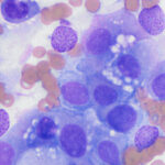
Examine the representative images of modified Wright-stained smears of the splenic aspirate (Figure 2), then answer the questions below:
- Based on the cells in the smears, what is your cytologic diagnosis?
- Do you think the mast cell tumor has metastasized to the spleen as well as the right medial iliac lymph node?
- Which additional tests would you perform to obtain a definitive diagnosis?
|
|
|
|


Answers on next page