
Peripheral blood and lymph node aspirates from a 12-year-old dog
Case Information
A 12-year-old male castrated Goldendoodle was presented to Cornell’s Small Animal Community Practice for a suspected seizure two weeks prior. The patient was otherwise well, was maintained on Nexgard® and Interceptor® as preventatives, and for the past year had been receiving monthly Librela™ (bedinvetmab, an anti-nerve growth factor antibody) injections for control of hip pain. No abnormalities were detected on physical examination, but blood was collected for a hemogram, with manual blood smear examination and differential cell count (Table 1, initial visit), and biochemical analysis. Results for the latter test were within reference intervals. The dog’s Librela™ injection was administered and the dog was sent home with plans to monitor for future seizure events. The patient was re-examined one month later due to another seizure; at this time the Librela™ and Nexgard® were discontinued, and the patient was started on a non-steroidal anti-inflammatory agent (meloxicam) for hip pain. A month later, another hemogram and biochemical panel was done to monitor for any side effects of prolonged meloxicam administration (Table 1, NSAID monitoring on second visit). The biochemical panel again did not reveal any abnormalities. The patient was returned two weeks later to Cornell University Hospital for Animals (CUHA) due to a third seizure and another hemogram was performed (Table 1, third seizure visit).

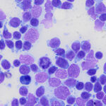
At this time, a biochemical panel revealed marginal hypoalbuminemia (3.1 g/dL, reference interval, 3.2-4.1 g/dL). Mild generalized peripheral lymphadenopathy was detected on physical examination and the left prescapular (measuring 1.6 x 2.6 x 3.0 cm) and popliteal (measuring 1.6 x 2.0 x 1.7 cm) lymph nodes were aspirated. Direct smears prepared from the aspirates were submitted for cytologic analysis.
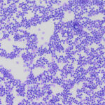
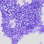
Examine the representative images of modified Wright-stained smears of the blood smear (Figures 1-2) and rapid-stained smears of the prescapular lymph node aspirate (Figure 3-5), then answer the questions below:
- Based on the hemogram data, clinical presentation, and images of the blood smear, what disease do you suspect in this patient??
- What additional tests could you pursue to further characterize the cells in the lymph node aspirates?
|
|
|
|
|
|
|


Answers on next page