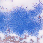
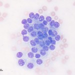
Photomicrographs of an aspirate from the right medial iliac lymph node from a dog
Case information
An 11-year-old female spayed Mastiff presented to the Cornell University Hospital for Animals with tenesmus (straining to defecate) over the past few months. The owners also reported that the dog’s stools were smaller than usual. The dog was otherwise active and eating normally with no coughing, sneezing, vomiting, diarrhea, polyuria or polydipsia. The referring veterinarian performed a complete blood count and serum chemistry, which revealed no abnormalities. On presentation, the dog was bright, alert, and responsive. The dog was normothermic (100.6°F), mildly tachypneic for her size (120 beats per minute), and was panting with no increased respiratory effort. Physical examination revealed moderate dental tartar, mild ceruminous discharge in both ears, bilateral pelvic limb muscular atrophy, an 8 cm left perianal mass, and several 2 cm skin masses on the right stifle, left tarsus, and dorsal aspect of neck. The remainder of the physical examination was unremarkable. No abnormalities were detected in a urinalysis, which was done to complete a minimum database. Thoracic radiographs were taken and showed no evidence of metastatic neoplasia. Abdominal ultrasonography was then performed, which revealed bilateraly enlarged medial iliac and inguinal lymph nodes. An ultrasound-guided fine needle aspirate of the right medial iliac lymph node was performed and submitted for cytologic evaluation (see images below):
- Are the cytologic findings expected from a lymph node aspirate? What cell type is used to confirm aspiration of a lymph node?
- How would you classify the dominant population of cells (epithelial, mesenchymal, or discrete cell)?
- Does this cell population appear well-differentiated and uniform or do the cells show cytologic criteria of malignancy?
|
|
|
Answer on next page


