
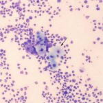
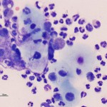
Photomicrographs of peritoneal fluid from a foal
Case information
A 2-day-old Thoroughbred colt presented to the Cornell University Equine and Farm Animal Hospital Emergency Service with the chief complaints of a weak suckle reflex and reluctance to rouse from sleep. On presentation the foal was bright, alert and responsive with mildly hyperemic mucous membranes and a normal capillary refill time (<2 seconds). His rectal temperature was 101.5°F (within normal limits for a foal). He was mildly tachypneic (respiratory rate of 50 breaths per minute). Several congenital abnormalities were noted including a fully reducible umbilical hernia, a scrotal hernia with scrotal and preputial edema, bilateral entropion, and mild flexural deformities of the forelimbs. The only abnormality detected on abdominal ultrasound examination was a small loop of intestine within the scrotal hernia. Initial bloodwork demonstrated a mild polycythemia (packed cell volume 48%, reference interval, 34-46%), increased total solids (7.9 g/dL, reference interval, 5.3-7.7 g/dL) and a lactic acidosis (blood lactate 2.9 mmol/L, reference interval, 0.3-1.5 mmol/L), which were attributed to dehydration and associated anaerobic metabolism. The foal was treated with intravenous fluids, broad-spectrum antibiotics and gastroprotectorants. The next day, the foal was bright and alert, but had increasing abdominal distension and a small amount of milk was observed coming out of the nostrils. At this time, a hemogram revealed an inflammatory leukogram, with a normal neutrophil count (2.8 x 10³/uL, reference interval, 2.8-6.6 x 10³/uL) and a mild left shift (1.8 x 10³/uL band neutrophils, reference interval 0 x 103/uL). The foal also had a hyperfibrinogenemia (400 mg/dL, reference interval 0-200 mg/dL). Abdominal fluid was collected for cytologic analysis. After viewing the photomicrographs of a direct smear of the abdominal fluid, consider the following questions:
- How would you categorize the effusion based on the cytologic findings?
- What is the origin and significance of the cells identified by the arrow in Figure 1? A closer view of the same cells is shown in Figure 2.
- What is the likely initiating cause of the foal’s problem?
|
|
|
Answer on next page