
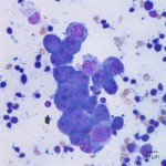
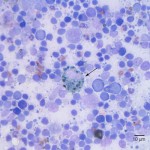
Photomicrographs of an aspirate from the right mandibular lymph node from a dog
Case information
A 14 year old male castrated Maltese presented for evaluation of an oral mass. Ten months previously, a mass was removed from the dog’s gingiva, which was diagnosed by histopathologic examination as a presumptive plasma cell tumor, that was incompleteley excised. Three months previously, a small mass was noted at the site of the previously excised tumor, which continued to grow. On the current physical examination, there were several proliferative coalescing masses on the buccal mucosa. They were predominantly on the right side of the mouth centered around the area of the previous biopsy, with several other raised masses on the right commissure. The dog had moderate dental tartar and gingivitis and prominent mandibular lymph nodes. Fine needle aspirate samples were taken from the right and left mandibular lymph nodes for cytologic evaluation and incisional biopsies were taken of two of the oral masses for histopathologic evaluation. Evaluate the representative photomicrographs (taken from two different slides prepared from the right mandibular lymph node aspirate) and consider the following questions:
- What are the potential lineages of the cohesive cells in Figure 1 and what is the significance of their presence in the lymph node?
- What is the pigmented cell indicated by the arrow in Figure 2 and what is its significance?
- How would you describe the lymphoid population in the sample?
|
|
|
Answer on next page