
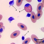
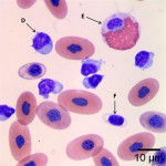
Photomicrographs of blood smear from a red-tail boa
Case information
A 5 year old male red-tail boa (boa constrictor) presented for weight loss. The snake had been completely inappetent for 6 to 7 months. The veterinarian submitted blood and feces to the Animal Health Diagnostic Center, associated with Cornell University, for a CBC and qualitative fecal exam (sugar flotation and zinc sulfate flotation). The fecal exam was negative. The most dramatic abnormality on the CBC was a marked lymphocytosis of 53.7 thousand/μl (reference interval 0.4-6.0 thousand/μL)1.
Evaluate the representative photomicrographs and consider the following:
- What morphologic features can be used to distinguish heterophils from eosinophils and lymphocytes from thrombocytes in non-mammalian blood?
- Identify the cells labeled A through F.
- What morphologic abnormality is present in the lymphocytes?
- Compare cells B and E. What is the significance of the cytoplasmic appearance of cell E?
|
|
|
Answer on next page
Pages: 1 2