
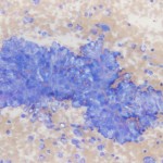
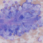
Photomicrographs of a fine needle aspirate of an adrenal mass in a ferret
Case information
The patient, a six year-old male castrated ferret, presented with a 1.5 month history of progressive, pruritic alopecia that started on the tail and spread to other areas of the body. The patient had a history of chronic pruritic seborrhea sicca, but it had worsened. Additionally, the owner had noticed recent polydipsia, but polyuria could not be ascertained.
He was bright, alert and responsive and in good body condition upon presentation. Physical examination revealed patchy, symmetrical alopecia along the dorsal midline, the entire ventrum, over the axilla, on all four paws and the entire tail. There were also three small circular eschars (3-4 mm diameter) on the ventrum and left hind paw, suspected to be evidence of self-trauma.
An abdominal ultrasound revealed a large (3 x 2.3 cm) mass in the area of the right adrenal gland, dorsal to the caudal vena cava, which had invaded the lumen of the caudal vena cava causing a thrombus to form. There was also a pocket of fluid associated with the mass that seemed to wrap dorsally and caudally around the right kidney. Ultrasound-guided needle aspiration of the mass was performed and samples submitted for cytologic analysis. Evaluate the representative photomicrographs below and consider the following questions:
- What is the cytologic diagnosis and underlying disease process?
- What other diagnostic tests would help confirm the diagnosis?
- What other clinical signs are common with this disease?
|
|
|
Answer on next page