
Bicavitary effusion in a cat
Case information
A 3 to 4 year old female spayed Maine Coon cat presented to the Cornell University Hospital for Animals Emergency Service for respiratory distress. The owner reported that she began hiding and acting unusual 3 days prior. The owner had also noted her having some increased respiratory effort. Over the next two days, she was hiding more, her appetite had decreased, and her respiratory effort had become markedly increased. She had a history of a heart murmur, but no other prior medical problems.
On presentation, she was in respiratory distress. The owner reported that she began hiding and acting unusual 3 days prior. The owner had also noted her having some increased respiratory effort. Over the next two days, she was hiding more, her appetite had decreased, and her respiratory effort had become markedly increased. She had a history of a heart murmur, but no other prior medical problems.
On presentation, she was in respiratory distress and immediately placed into an oxygen cage. After an intravenous catheter was placed, she was given a mild sedative intravenously and a FAST ultrasound scan was performed. The FAST scan revealed the presence of pleural and pericardial effusion. She was given additional sedatives to facilitate a thoracocentesis and pericardiocentesis. The thoracocentesis yielded 190mL of orange-tinged serous fluid and the pericardiocentesis yielded 140mL of serosanguineous fluid. The pleural and pericardial fluids were submitted to the Clinical Pathology laboratory for fluid analysis and cytology.
The fluid analyses and representative cytologic images from both sites are provided below:
| Site of effusion |
Pleural
|
Pericardial
|
| Volume (mL) |
3.0
|
4.0
|
| Color |
Medium orange
|
Dark red
|
| Turbidity |
Slightly cloudy
|
Opaque
|
| Total protein-refractometer (g/dL) |
2.8
|
4.3
|
| Nucleated cells (thou/μL) |
9.3
|
32.5
|
| Red blood cells (thou/μL) |
22.3
|
1826.7
|
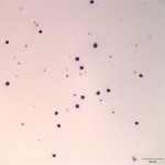
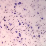
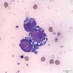
View the photomicrographs then answer the questions below.
- What is the predominant cell type in the pleural fluid?
- How would you classify the pleural effusion?
- How do the cells in the pericardial fluid differ from those in the pleural fluid?
- What is your interpretation of the pericardial fluid?
|
|
|
|
Answer on next page