
Peripheral blood from a cat
Case information
Whole blood from a 3 year old male neutered Himalayan cat was collected into an EDTA tube and submitted to the Animal Health Diagnostic Center at Cornell University for a complete blood count. The submitting hospital provided no clinical history.
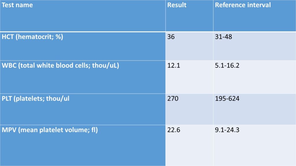
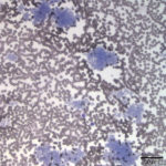
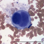
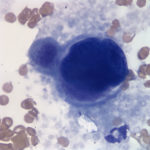
Review the pertinent data generated by the automated hematology analyzer (ADVIA 2120) and evaluate the photomicrographs below to answer the following questions:
Complete Blood Count (CBC) – Truncated; Data from analyzer
Questions:
- For which cell line is the analyzer clearly reporting spuriously decreased results?
- What is an artifact on a typical serum biochemistry panel that may be associated with this condition (in dogs), and what other type of specimen could you evaluate to confirm the artifact?
- How would you classify the large cells in Figures 2-3?
|
|
|
|
Answers on next page.
Pages: 1 2