
Tracheal wash from a horse
Case Information
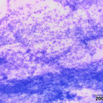
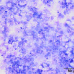
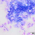
A 14 year old Paint mare was referred to the Cornell Equine Nemo Farm Animal Hospital with a one week history of decreased appetite and a 2-day history of pipestream diarrhea and fever, leading to a preliminary diagnosis of colitis. The day of referral, the referring veterinarian had documented a fever, cold extremities, cyanosis, tachypnea, and tachycardia. Normal gut sounds were auscultated. On arrival at Cornell, physical examination revealed fever, dehydration (prolonged skin tent and capillary refill time > 3 seconds), toxic mucosa, injected sclera, colic associated with passing watery diarrhea, tachypnea and tachycardia. Point-of-care bloodwork revealed a moderate erythrocytosis (59% packed cell volume, reference interval [RI]: 31-48%), severe hyponatremia (118 mEq/L, RI: 134-142 mEq/L), moderate to severe azotemia (5.6 mg/dL creatinine, RI: 0.8-1.5 mg/dL) and lactic acidosis (6.9 mmol/L, normal <2.0 mmol/L). A hemogram performed the next day showed a neutropenia (2.0 x 10³/μL, RI: 2.7-6.6 x 10³/μL) with a mild to moderate left shift (1.3 x 10³/μL, RI: 0-0.1 x 10³/μL) and marked toxic change in neutrophils. The fibrinogen concentration by heat precipitation was mildly increased at 400 mg/dL (RI: 0-200 mg/dL). Thoracic auscultation revealed harsh lung sounds. A tracheal wash was collected via an endoscope and submitted for cytologic analysis. Examine the images below then answer the provided questions.
- What abnormal findings are present in the tracheal wash?
- What is your cytologic diagnosis?
|
|
|
|
|

Answers on next page