
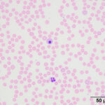
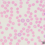
Venous blood smear from a dog
Case Information
An 11-year-old female spayed Doberman Pinscher was presented to the Cornell University Hospital for Animals with a 24-hour history of reduced appetite. The dog had also urinated in the bed the previous evening. Six weeks prior, the dog had been admitted to the clinic as an emergency for an acute onset of vomiting and abdominal pain. An exploratory laparotomy was performed, which revealed abdominal adhesions and a gastric perforation. About 2/3 of the stomach was resected and a splenectomy was concurrently performed. The dog had previously tested positive for von Willebrand Disease (vWD; von Willebrand factor antigen concentration: 16%, reference interval, 70-180%) 2 years prior and was given blood transfusions during surgery to assist with surgical hemostasis. Histopathologic evaluation of the resected stomach wall revealed gastritis with Helicobacter organisms and a chronic peritonitis.
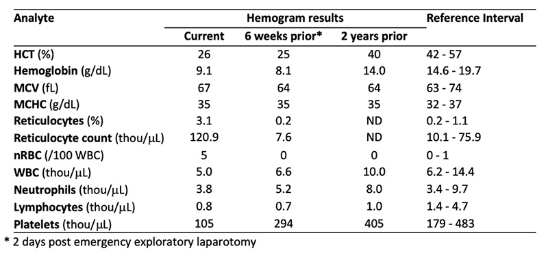
On physical examination, the dog had a normal heart and respiratory rate and was not febrile. No abnormalities were detected on abdominal palpation or thoracic auscultation. Blood was taken for a hemogram and biochemical panel. The biochemical profile revealed no relevant abnormalities. Hemogram results are shown below, along with previous results that were taken 2 days after the exploratory laparotomy and 2 years earlier (when vWD testing was done). A hemogram was not done before the emergency surgery. View the hemogram results and representative images of the blood smear (Figures 1-2) then answer the questions below.
- What is your diagnosis from the blood smear?
- What factors would have predisposed the dog to this condition?
|
|
|
Answers on next page