
Venous blood from a cat
Case Information
A 1 year old neutered male domestic shorthair cat was presented to a veterinarian for the primary complaints of chronic upper respiratory symptoms, lethargy, and decreased appetite. The cat was documented to be negative for feline leukemia virus (FeLV) and feline immunodeficiency virus (FIV) via a rapid test performed at a humane shelter. Upon physical examination, the cat was febrile (104.4°F) and lethargic with bilateral conjunctivitis, prolapsed third eyelids, upper airway congestion and a mucopurulent nasal discharge. The cat was treated with subcutaneous fluids, antibiotics, an antiviral medication (famcyclovir), and an anti-inflammatory medication. The patient showed mild improvement with treatment but symptoms recurred 4 days after the initial examination. A complete blood count (CBC), with an automated white blood cell differential count, performed at the veterinary clinic showed a low normal erythron (hematocrit [HCT], 32%, reference interval [RI], 30-52%) with a marked leukocytosis (218.9 x 10³/µL, RI, 2.9-17.0 x 10³/µL) characterized by a marked monocytosis (124.6 x 10³/µL, RI, 0.1-0.7 x 10³/µL) and lymphocytosis (93.6 x 10³/µL, RI, 0.9-6.9 x 10³/µL). There was a concurrent marked neutropenia (0.5 x 10³/µL, RI, 2.3-10.3 x 10³/µL) and moderate thrombocytopenia (78 x 103/µL, RI, 151-600 x 103/µL).
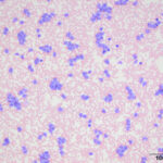
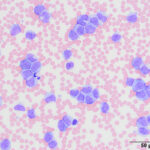
The veterinarian submitted an EDTA-anticoagulated blood sample and an unstained blood smear to the Clinical Pathology laboratory in the Animal Health Diagnostic Center at Cornell University for evaluation by a clinical pathologist. On receipt (24 hours after collection), the blood was analyzed with an automated hematology analyzer (Table 1) and a blood smear examination with manual differential count was performed on the submitted blood smear, which was stained with a modified Wright’s stain (Table 1, Figures 1 -3).
- What is your diagnosis?
- What diagnostic tests can be used to further characterize the cells in blood?
|
|
|
|
Answers on next page