
Aspirate from a left axillary mass in an alpaca
Case Information
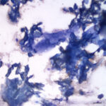
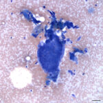
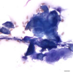
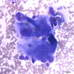
An 11-year-old male alpaca was presented to a veterinarian for a mass in the left axilla. The mass had been discovered one year prior during shearing and was approximately 1 x 2 cm and firm on palpation. The mass had grown substantially in the intervening year and was approximately 6 x 6 x 6 cm when examined by the attending veterinarian. No abnormalities were noted in the overlying skin or hair fibers. The mass was non-painful on palpation and appeared cystic superficially with a firmer deep portion that was well-attached to underlying tissues. Aspirates were taken from the superficial and deep portions of the mass and submitted to the Animal Health Diagnostic Center at Cornell University for cytologic evaluation (Figures 1-4). No digital slide is available for this case.
- What cell types can you identify in the aspirate?
- What are your differential diagnoses for the mass?
|
|
|
|
|
Answers on next page