
Chronic uveitis in a dog
Case information
A 3 year old spayed female Pomeranian was presented for a 7 month history of conjunctival hyperemia and blepharospasm in the right eye (OD) that improved after topical corticosteroid treatment, but recurred within weeks of discontinuation. On ophthalmologic examination, the right eye had conjunctival hyperemia, diffuse corneal edema, ciliary flush, trace aqueous flare, anterior chamber fibrin, iris hyperpigmentation, miosis, and a mature intumescent cataract. Increased intraocular pressure was detected (40 mmHg). A moderate leukocytosis (24.5 x 10³/μL, reference interval, 5.7-14.2 x 10³/μL) attributable to a mature neutrophilia was present on a hemogram. Serum biochemistry panel and urinalysis were unremarkable. Ultrasonographic examination of the right eye revealed complete bullous retinal detachment with diffuse vitreal and subretinal opacification. Urine, ocular, and blood aerobic bacterial, mycoplasmal, and fungal cultures were submitted. Pending results, topical treatment with broad-spectrum antibiotics and anti-inflammatory corticosteroids (neomycin-polymyxin β-dexamethasone ointment) and anti-glaucoma agents (dorzolamide-timolol solution) was initiated.
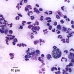
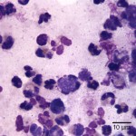
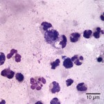
Three days after initial presentation, the dog presented with hypopyon and severe ocular discomfort in the right eye. Aqueous aspirates were performed under sedation. Cytologic evaluation of all samples revealed similar findings; representative cytologic images are provided below. After examining the images, answer the questions posed below:
- What is your diagnosis based on the cytologic findings?
- What are two differential diagnoses for the causative agent?
- What additional tests could be performed?
|
|
|
|
Answer on next page.