
Tracheal wash from a cow
Case Information
Approximately 90% of the cows in a herd of 65-70 Holstein cattle in upstate New York were affected with an acute onset of severe coughing and dyspnea, which rapidly spread through the herd. Many of the cows were treated with penicillin G followed by ceftiofur antibiotics for presumptive bacterial pneumonia, with minimal improvement. When the submitting veterinarian visited the farm, a small subset of affected cows was examined. About half of the cows were febrile, while the other half were normothermic. On auscultation, the cows’ lungs were wet and raspy with a generalized distribution. A transtracheal wash was performed on a 3 year-old female Holstein cow, selected as a representative animal. The acquired tracheal wash sample was submitted to the Animal Health Diagnostic Center for cytologic evaluation, as well as aerobic bacterial culture and sensitivity, and virologic testing.
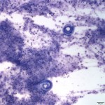
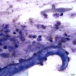
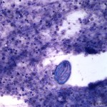
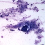
Direct and sediment smears were prepared from the submitted fluid. Representative images from the Wright’s stained sediment smears are present below. After examining the images, answer the following questions:
- How would you classify the inflammation present?
- What infectious agent is the cause of the inflammation and clinical signs in this cow?
- What other diagnostic test can be done to diagnose this infection?
|
|
|
|
|
Answer on next page