
Blood and bone marrow smears from a steer
Case information
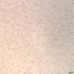
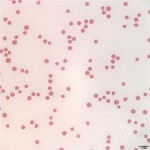
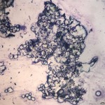
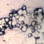
An 8 month old Holstein steer presented with bloat and lethargy. On physical examination, there were petechial hemorrhages on the gums and tongue. Abnormal eructation sounds were noted during the examination. A CBC was performed with results summarized below (Table 1), including images from the blood smear (Figure 1a & 1b). Four days later, the steer died. On field necropsy there was diffuse petechiation. No large hemorrhages were noted. Impression smears from the bone marrow were collected immediately post-mortem (images below, Figure 2a & 2b). Four more live cattle on the farm were found to have petechial hemorrhages. One was necropsied, with sections from the stomach and ileum showing moderate to severe, diffuse, acute, submucosal hemorrhage on histopathologic examination. The cattle are located in a large fenced area of clear-cut woodland and fair-quality hay was available free-choice.
Evaluate the CBC results and representative photomicrographs of blood and bone marrow below and consider the following questions:
- What single word can be used to summarize the CBC findings? Does the anemia appear regenerative or non-regenerative?
- What abnormality is noted in the bone marrow? How do the CBC and bone marrow findings relate to the steer’s petechiation?
- What is the primary differential diagnosis for these CBC and bone marrow findings in cattle?
| Table 1: Abbreviated test results | |||
| Test | Results | Units | Reference interval |
| Hct | 11 L | % | 25-33 |
| Hgb | 3.7 L | g/dL | 8.7-12.4 |
| RBC | 2.9 L | mill/μL | 5.0-7.2 |
| MCV | 37 L | fl | 38-51 |
| MCH | 13 L | pg | 14-19 |
| MCHC | 35 | g/dL | 34-38 |
| WBC | 2.1 L | thous/μL | 5.9-14.0 |
| PMN | 0 | thous/μL | 1.8-7.2 |
| LYM | 2.1 | thous/μL | 1.7-7.5 |
| PLAT | <15 L | thous/μL | 252-724 |
|
|
|
|
|
Answer on next page