
Nasal mass in a cat
Case information
A 12 year-old, female spayed, domestic shorthair cat was presented to the Cornell University Hospital for Animals for a recheck examination for worsening nasal congestion. The cat historically had episodic nasal congestion for 3 years, which responded to treatment with azithromycin. Over the past seven months, the congestion became less responsive to azithromycin and prednisone. On presentation, the cat was quiet, alert and responsive and had a mildly increased rectal temperature (102.7°F), but all other vital parameters were within normal limits. The cat had audible nasal stertor with inspiratory and expiratory effort and was open-mouth breathing. Blood samples were obtained and QATS (quick assessment tests) revealed a normal packed cell volume of 35%, increased total solids of 8.8 g/dL, a mild azotemia (urea nitrogen of 15-26 mg/dL) and normal glucose values (142 mg/dL). A computed tomography (CT) scan was performed and revealed a nasal mass with fluid in the left nasal cavity and sinuses. There was minimal bony lysis of the nasal turbinate. The mass was partially removed and touch impression smears of the mass were submitted for cytological evaluation. The remaining tissues were submitted for histopathologic examination.
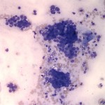
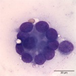
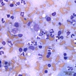
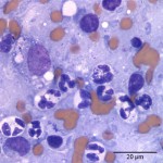
Evaluate the following representative photomicrographs of the mass impression smears (Figures 1-4) and answer the following questions:
- How would you classify the large cell population (epithelial, mesenchymal, round cell)?
- Is there evidence of inflammation and an infectious agent?
- What is your cytologic diagnosis?
|
|
|
|
|
Answer on next page