
Perilimbal mass in a horse
Case information
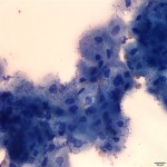
A 6 year-old male castrated Quarter Horse from Montana was presented to the submitting veterinarian in Kansas for a mass on the right eye that the owner believed had been present since the horse was purchased eight months ago. On the right eye, the mass arose from the ventromedial perilimbal conjunctiva from 3-5 o’clock and invaded the adjacent cornea. The mass was diffuse and flat with a roughened surface.
Impression smears were made from the biopsies taken by the veterinarian. The smears and biopsied tissue were submitted to the Animal Health Diagnostic Center for cytology and histopathology, respectively.
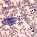
Evaluate the representative Wright’s stained cytology images below and consider the following questions:
- How would you classify the cell population in Figure 1 (epithelial, mesenchymal, round cell)?
- a) What does the cytoplasm color indicate about the cells present in Figure 1?
b) Why is this abnormal (given the location of the impression smear)? - What are the cells indicated by the arrows in Figure 2?
- How would you interpret the bacteria present in the sample?
|
|
|
Answer on next page