
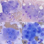
Photomicrographs of an aspirate from a liver mass in a dog
Case information
A 5 year old, spayed female mixed breed dog presented to the Cornell University Hospital for Animals (CUHA) for removal of a cutaneous mast cell tumor. Upon physical examination, additional cutaneous tumors were noted and sampled via fine-needle aspiration. The cytologic results were consistent with mast cell tumors. A full diagnostic evaluation was performed by the CUHA oncology service. A complete blood count revealed no hematological abnormalities. On serum chemistry analysis, the following abnormalities were noted: Panhyperproteinemia (albumin: 8.1 g/dL, reference interval, 3.1-4.1 g/dL; globulins: 3.9 g/dL, reference interval, 1.9-3.6 g/dL), mild hypoglycemia (57 mg/dL, reference interval, 60-120 mg/dL), and mild to moderate increases in hepatic enzyme activities (ALT: 2118 U/L, reference interval, 25-106 U/L; AST: 168 U/L, reference interval, 16-50 U/L; ALP 284 U/L, reference interval, 12-122 U/L). An abdominal ultrasound revealed a large (8.4 x 7.7 x 6.5 cm) contrast-enhancing mass in the right lateral lobe of the liver. The mass was aspirated with ultrasound guidance and the specimen was submitted for cytological analysis. Examine the image compilation below then answer the following questions:
- What is your cytologic diagnosis?
- What is a potential mechanism for the mild hypoglycemia?
- What is a potential explanation for the marked hyperalbuminemia?
Answer on next page