
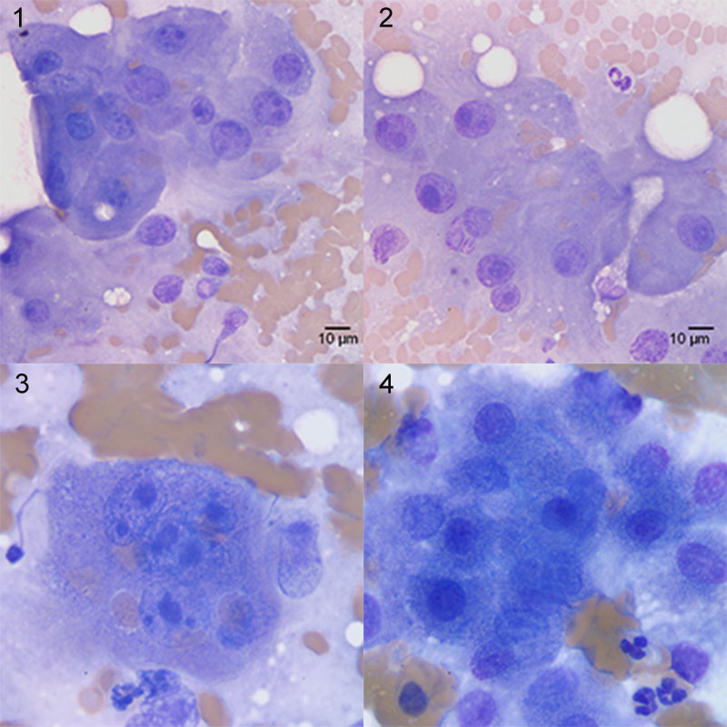
Figure: Images 1 through 3 are representative of the aspirate of the dog’s liver mass (Wright’s stain). Image 4 represents an aspirate from the liver of a different dog for comparison (taken at the same magnification). The liver of the latter dog was aspirated for staging of lymphoma (Wright’s stain).