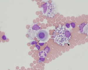
A sample of peritoneal fluid from a dog with congestive heart failure was lightly red and cloudy with a nucleated cell count of 2,500 cells/uL and a RBC count of 120,000/uL. The total protein by refractometer was 3.0 g/dL. Cytospin smears of the fluid contained a mixture of neutrophils (non-degenerate) and vacuolated and phagocytic macrophages, with fewer small lymphocytes. Plasma cells were also seen (indicating localized antigenic stimulation, not shown) as were isolated mesothelial cells (arrow). Macrophages contained hemosiderin and erythrocytes (erythrophage, arrowhead) in their cytoplasm. The latter finding with the lack of platelets and presence of high numbers of red blood cells in the background indicate concurrent diapedesis of red blood cells or hemorrhage into the abdominal cavity. Congestive heart failure is a common cause of this type of abdominal effusion in dogs. Wright’s stain, 500x magnification.

