
Interpretation
Moderate mixed, mostly neutrophilic, inflammation with “ragocytes” and “lupus erythematosus (LE)” cells, indicating immune-mediated joint disease
Explanation
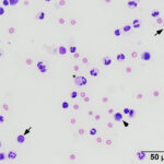
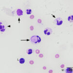
Direct and sediment smears were highly cellular, consisting of 74% mildly degenerate neutrophils, 21% lymphocytes (often called “small mononuclear” cells), 4% macrophages or synovial lining cells (often called “large mononuclear” cells), and 1% eosinophils with a small amount of blood. The predominance of neutrophils in a highly cellular sample confirms an inflammatory process (Question 1). Many neutrophils contained variably-sized purple globules in their cytoplasm; these cells are often termed “ragocytes” or “ragocyte-like” cells.1 Rare neutrophils contained a single, large, round, smooth, pink inclusion in the cytoplasm, which displaced the nucleus to the periphery of the cell; these are known as LE cells2 (Figures 1-2). Leukophagia was noted in macrophages and a few reactive lymphocytes, including large forms, were seen. Cells were windrowing minimally, fitting with the visual estimate of decreased viscosity. No infectious organisms were identified. The granular and large inclusions in “ragocyte-like” cells and LE cells are thought to represent phagocytized immune complexes and nuclear material, respectively.2,3 The presence of both these cells favors an immune-mediated pathogenesis for the polyarthropathy and the LE cells, in particular, are supportive of a diagnosis of systemic lupus erythematosus (SLE)4 (Question 2).
|
|
|
Outcome
The dog responded well to a tapering course of corticosteroids, but symptoms recrudesced some time later. Another tapering course of corticosteroids was initiated, to which the dog responded well.
Discussion
Immune-mediated polyarthropathy (IMPA) is a broad category of joint disease encompassing multiple causes in the dog. IMPAs can be primary, due to an auto-immune dysregulation with no initiating cause, or secondary to an inflammatory process distant from the joints. The suggested mechanism for this process is a type III hypersensitivity reaction wherein antigen-antibody complexes deposited within the joint space drive complement fixation and thus local inflammation.5 Secondary IMPAs may be attributed to a various causes, including infectious and inflammatory disease. An important class of infectious agents associated with secondary IMPA in the northeastern part of the US are tick-borne diseases (e.g., borreliosis, anaplasmosis, ehrlichiosis). Other infectious causes such as leptospirosis have been reported,6 and we theorize that other, as yet unrecognized, infectious organisms could drive an IMPA. Noninfectious inflammatory processes, such as gastroenteropathy,7 drugs and vaccines,8,9 and neoplasia,7 are associated with IMPA. Secondary IMPAs are generally diagnosed by serologic testing for a suspected infectious agent, uncovering and treating another inflammatory (e.g., an enteropathy) or neoplastic process, or having a history of recent pharmaceutical administration or vaccination.
Primary IMPAs can be divided into erosive or non-erosive forms based on evidence of bone destruction on radiographs.10 Non-erosive primary IMPA causes include SLE and breed-associated arthropathies, such as Shar-Pei fever, or are idiopathic. Erosive primary IMPAs include rheumatoid arthritis (RA), and breed-associated arthropathies, such as Border Collies with trapped neutrophil syndrome.11 Diagnostic confirmation of primary IMPAs is challenging and often predicated on excluding secondary causes and monitoring response to treatment. Ancillary testing for antinuclear antibodies (ANA) can be done, as discussed below, but the test still must be interpreted in light of clinical signs and the results of other diagnostic testing.
SLE is an auto-immune multisystemic disease driven by anti-nuclear antibodies, which manifests with various clinical signs. IMPA is the most common clinical presenting sign of SLE in dogs, but the disease can also result in a hemolytic anemia, vasculitis, dermatologic lesions, or glomerulonephropathy.12 Given the highly diverse presentation of SLE in both humans13 and dogs,12 definition of the disease and diagnostic criteria are broad and periodically under revision in humans. The most recent criteria in humans, generated by the European League Against Rheumatism and the American College of Rheumatology, requires an ANA titer of 1:80 as an entry criterion, and following that, a score of >10 accumulated by weighing clinical signs (e.g., fever, psychiatric signs, ulcers, or cavitary effusion, among many others) and supportive diagnostic testing (e.g. low complement concentrations, the presence of specific autoantibodies, or a renal biopsy supportive of lupus nephritis).13 There is currently a lack of consensus on establishing a diagnosis of SLE in the dog,12,14,15 but all authors agree on the utility of multiple findings, including clinical signs (e.g., fever, or dermatologic disease), and positive ANA titers. Notably, a positive ANA titer may be present in dogs that are healthy12,16 or have other inflammatory conditions (e.g., leishmaniasis).16 Broadly, healthy dogs and dogs with inflammatory disease tend to have lower ANA titers (<1:256) than those with SLE, however markedly high titers (≥1:4096) have been reported in both groups.16 Additionally, there are human cases with negative ANA titers but other features suggestive of SLE.13,17 However, persistently negative ANA titers are a contentious point in SLE diagnosis in humans and an exclusion criteria by the current classification guideline noted above.13 This data underscores the difficulty of conclusively diagnosing SLE even in humans, where there is access to a greater barrage of diagnostic tests than we have available in dogs. This is all to say that ANA testing does not rule in or out SLE as definitively as one would hope.
Cytologic assessment of joint fluid in IMPA generally reveals neutrophilic inflammation regardless of inciting cause. Neutrophils are typically not degenerate and contain no cytoplasmic inclusions. The cytologic findings in this case depart from the usual expectations of a standard IMPA given the presence of “ragocyte-like” cells and LE cells. The nature of ragocytes is incompletely explored in veterinary medicine and some authors contest the use of the term in stained smear preparations,18 hence the term “ragocyte-like” used herein. Indeed, the human literature generally uses the term only in the context of unstained wet-mount preparations19 with the original paper going so far as to claim these inclusions do not stain with Wright- or Giemsa-based stains.20 Current opinions favor that the purple granules in the cytoplasm of the neutrophils represents phagocytized immune complexes.1 Generally, ragocytes are accepted to be suggestive of, but not pathognomonic for, immune-mediated disease in the veterinary literature,3 although they have been described in non-immune mediated conditions in humans.21 The paucity of investigations into their prevalence in veterinary medicine as well as the lack of concordance among human and veterinary descriptions prevents a more confident claim to their significance. LE cells are more thoroughly examined in the literature and have been shown to contain nuclear material that has been ingested by neutrophils,2 a process driven by the presence of ANA, such as those in SLE. Specific preparations to elicit LE cells, such as the LE test, have fallen out of practice in human medicine in favor of less laborious testing, such as specific autoantibody titers and complement levels,13,22 and the LE test was removed as a human SLE diagnostic criterion in 1997.23 Although this test is highly specific for SLE (although not without false positives in rheumatoid arthritis and potentially other immune-mediated syndromes24), a poor sensitivity has relegated this test to the annals of history in human medicine.25 Similar to the findings in humans, the LE cell preparation is of limited sensitivity on dogs,26 but specificity remains high, with rare positive results being reported in dogs with lymphoid neoplasia and dicoumarol poisoning.27 However, the likelihood of finding a laboratory still performing this test is slim. Due to evolving disease definitions and a general dearth of literature on canine SLE, the true prevalence of LE cells in vivo in dogs has not established. However, finding these cells in vivo, usually in joint fluid, appears to be highly suggestive of SLE.4 In the case herein, we used LE cells to indicate that it supported a diagnosis of SLE, but we lacked additional diagnostic criteria (e.g. evidence of a hemolytic anemia or glomerulopathy) to confirm SLE as the ultimate diagnosis.
Author: K Marciano (clinical pathology resident), edited by T. Stokol
References
- Camus MS, Almy FS, Cienava EA. A ragtime joint. Vet Clin Pathol. 2010;39(1):4–4. doi:10.1111/j.1939-165X.2010.00222.x
- Lee SL, Michael SR, Vural IL. The L.E. (Lupus erythematosus) cell: Clinical and chemical studies. Am J Med. 1951 Apr 1;10(4):446–51. doi:10.1016/0002-9343(51)90289-6
- Meléndez-Lazo A, Fernández M, Solano-Gallego L, Pastor J. What is your diagnosis? Synovial fluid from a dog. Vet Clin Pathol. 2015;44(2):329–30. doi:10.1111/vcp.12232
- Ningombam A, Sarkar A, Acharya S, Chopra A. Lupus erythematosus cell in body fluids: A case report and review of literature. Diagn Cytopathol. 2020;48(8):773–7. doi:10.1002/dc.24450/li>
- Kohn B. Canine immune-mediated polyarthritis. Eur J Companion Anim Pract. 2007 Oct;17(2):119–24/
- Furlanello T, Reale I. First description of reactive arthritis secondary to leptospirosis in a dog. Iran J Vet Res. 2020;21(2):146–9. PubMed PMID: 32849896; PubMed Central PMCID: PMC7430365.
- Bennett D. Immune-based non-erosive inflammatory joint disease of the dog. 3. Canine idiopathic polyarthritis. J Small Anim Pract. 1987;28(10):909–28. doi:10.1111/j.1748-5827.1987.tb01316.x
- Giger U, Werner LL, Millichamp NJ, Gorman NT. Sulfadiazine-induced allergy in six Doberman Pinschers. J Am Vet Med Assoc. 1985;186(5):479–84. doi:10.2460/javma.1985.186.05.479
- Kohn B, Garner M, Lübke S, Schmidt MFG, Bennett D, Brunnberg L. Polyarthritis following vaccination in four dogs. Vet Comp Orthop Traumatol. 2003;16(01):06–10. doi:10.1055/s-0038-1632747
- Johnson KC, Mackin A. Canine Immune-Mediated Polyarthritis Part 1: Pathophysiology. J Am Anim Hosp Assoc. 2012 Jan 1;48(1):12–7. doi:10.5326/JAAHA-MS-5744
- Hegler AK, Grooters AM, Dehghanpir SD, Gallaher RA, Gaschen LE. Trapped Neutrophil Syndrome in a Border Collie. J Am Anim Hosp Assoc.2 020;56(3):e563. doi:10.5326/JAAHA-MS-6981
- Fournel C, Chabanne L, Caux C, Faure JR, Rigal D, Magnol JP, et al. Canine Systemic Lupus Erythematosus. I: A Study of 75 Cases. Lupus. 1992 May 1;1(3):133–9. doi:10.1177/096120339200100303
- Aringer M, Costenbader KH, Daikh DI, Brinks R, Mosca M, Ramsey-Goldman R, et al. 2019 EULAR/ACR Classification Criteria for Systemic Lupus Erythematosus. Arthritis Rheumatol Hoboken NJ. 2019 Sep;71(9):1400–12. doi:10.1002/art.40930 PubMed PMID: 31385462; PubMed Central PMCID: PMC6827566.
- Rondeau MP, Walton RM, Bissett S, Drobatz KJ, Washabau RJ. Suppurative, Nonseptic Polyarthropathy in Dogs. J Vet Intern Med. 2005;19(5):654–62. doi:10.1111/j.1939-1676.2005.tb02743.x
- Gorman NT, Werner LL. Immune-mediated diseases of the dog and cat. I. Basic concepts and the systemic immune-mediated diseases. Br Vet J. 1986 Sep 1;142(5):395–402. doi:10.1016/0007-1935(86)90040-0
- Monier JC, Ritter J, Caux C, Chabanne L, Fournel C, Venet C, et al. Canine Systemic Lupus Erythematosus. II: Antinuclear Antibodies. Lupus. 1992 Oct 1;1(5):287–93. doi:10.1177/0961203392001005031
- Wallace DJ, Metzger AL. Positive LE preps and negative ANA in brothers with SLE: is there still a role for the LE cell prep? A literature review. J Rheumatol. 1992 Mar;19(3):497.
- Valenciano AC, Cowell RL. Cowell and Tyler’s Diagnostic Cytology and Hematology of the Dog and Cat. 5th ed. St. Louis, MO: Elsevier; 2020. p 556 .
- Freemont AJ. Microscopic analysis of synovial fluid–the perfect diagnostic test? Ann Rheum Dis. 1996 Oct;55(10):695–7. doi:10.1136/ard.55.10.695 PubMed PMID: 8984932; PubMed Central PMCID: PMC1010283.
- Hollander JL, McCarty DJ, Astorga G, Castro-Murillo E. Studies on the Pathogenesis of Rheumatoid Joint Inflammation I. The “R.A. Cell” and a Working Hypothesis. Ann Intern Med. 1965 Feb;62:271–80.
- Cats A, Lafeber GJ, Klein F. Immunoglobulin phagocytosis by granulocytes from sera and synovial fluids in various rheumatoid and nonrheumatoid diseases. Ann Rheum Dis. 1975 Apr 1;34(2):146–55. doi:10.1136/ard.34.2.146
- Steensma DP. Fifty Years of Tart Cells. Mayo Clin Proc. 1999 Sep;74(9):936–8. doi:10.4065/74.9.936
- Hochberg MC. Updating the American College of Rheumatology revised criteria for the classification of systemic lupus erythematosus. Arthritis Rheum. 1997 Sep 1;40(9):1725. doi:10.1002/art.1780400928 PubMed PMID: 9324032.
- Stephen R. Koller, Charles L. Johnston, Jr., Charles W. Moncure, Marion V. Waller. Lupus erythematosus cell preparation-antinuclear factor incongruity. A review of diagnostic tests for systemic lupus erythematosus. 1976 Sep;66(3):495–505.
- Tan EM, Cohen AS, Fries JF, Masi AT, Mcshane DJ, Rothfield NF, et al. The 1982 revised criteria for the classification of systemic lupus erythematosus. Arthritis Rheum. 1982;25(11):1271–7. doi:10.1002/art.1780251101
- Bennett D, Kirkham D. The laboratory identification of serum antinuclear antibody in the dog. J Comp Pathol. 1987 Sep 1;97(5):523–39. doi:10.1016/0021-9975(87)90003-X
- Lewis RM. Clinical Evaluation of the Lupus Erythematosus Cell Phenomenon in Dogs. J Am Vet Med Assoc. 1965 Jan;147(9):939–42.
- Braman SL, Bessett JP, Anderson KL, Friedrichs KR. Identification of Lupus Erythematosus Cells and Ragocytes in Peripheral Blood from a Dog with Systemic Lupus Erythematosus. Vet Clin Pathol. 2026 May 6, ePub ahead of print. doi:10.1111/vcp.70087
- Black LJ, Hechler AC, Duffy ME, Beatty SSK. Presumed lupus erythematosus cells identified in bronchoalveolar lavage fluid from a Mexican Hairless dog. Vet Clin Pathol. 2017;46(2):354–9. doi:10.1111/vcp.12471