
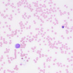
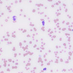
Venous blood smear from a horse
Case Information
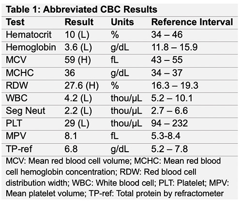
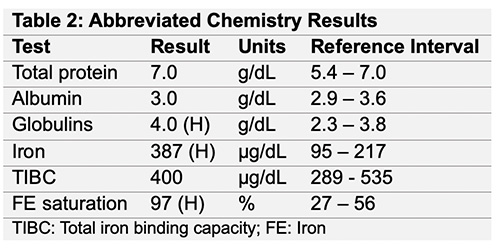
A 2-year-old Quarter Horse gelding was presented to the primary veterinarian for a 1-month history of progressive lethargy and decreased performance. The horse had no prior health issues. The primary veterinarian performed bloodwork, which revealed pancytopenia and the horse was subsequently referred to the Equine and Food Animal Hospital at Cornell University as an emergency the same day. On presentation, the horse was tachycardic and tachypneic, and had pale mucous membranes. A repeat hemogram confirmed pancytopenia (Table 1). A fibrinogen by heat precipitation was normal (200 mg/dL, reference interval, 0-200 mg/dL). A large animal chemistry panel showed mild abnormalities in globulins and serum iron concentration and percentage saturation. A urine dipstick on a voided urine sample done at point-of-care showed no abnormalities. Thoracic and abdominal ultrasonographic examination was performed and results were unremarkable.
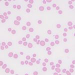
Representative images from the modified Wright’s-stained peripheral blood smear are shown below (Figures 1-3). After examining the images, answer the following questions:
- What abnormalities are evident in the blood smear?
- Do you think the severe anemia is regenerative based on the changes in the red blood cells?
- What is your top differential diagnosis for the pancytopenia?
|
|
|
|
Answers on next page