
Peritoneal fluid from a dog
Case Information
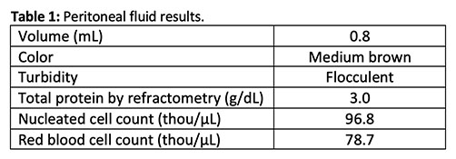
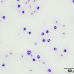
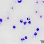
An 11 year-old, female spayed, mixed breed dog was presented to Cornell University Hospital for Animals (CUHA) for a suspected septic peritonitis. The patient had been seen for repeated visits by the referring veterinarian (RDVM), over a 4 day period, for persistent and progressive lethargy, discomfort, and anorexia. The patient was initially treated with Deramaxx® (deracoxib), an oral non-steroidal anti-inflammatory drug (NSAID), for pain from a suspected injury. The next day, the patient was represented to the RDVM after vomiting and acting abnormally at home. The patient was hospitalized and a complete blood count (CBC), biochemical panel, and pancreatic lipase snap test were performed. The CBC showed a marked leukocytosis (50.0 thou/µL; reference interval [RI]: 6.0-17.0 thou/µL), characterized by a marked neutrophilia (38.7 thou/µL; RI: 3.0-12.0 thou/ µL), a left shift with no indication of the degree of toxic change (band neutrophils, 3.8 thou/µL; RI: 0-0.1 thou/µL), a moderate monocytosis (4.2 thou/µL; RI: 0.2-1.5 thou/µL), and a marked thrombocytopenia (14 thou/µL; RI: 165-500 thou/µL). The biochemical panel revealed a mild hypoalbuminemia (2.2 g/dL; RI: 2.5-4.4 g/dL), hyperphosphatemia (7.8 mg/dL; RI: 2.9-6.6 mg/dL), and azotemia (urea nitrogen [UN], 24 mg/dL; RI: 7-25 mg/dL; creatinine, 1.9 mg/dL: RI: 0.3-1.4 mg/dL). The pancreatic lipase snap test was negative. The patient was started on antibiotics and given a NSAID injection (Meloxicam). The patient continued to do poorly and was hospitalized the next day for intravenous fluid therapy, and injectable antibiotic, pain, and dexamethasone treatment. An abdominal radiograph was performed, which revealed a possible foreign body in the cranial abdomen and an abnormal gastric profile. A barium study was performed, which showed contrast passing through the stomach and into the duodenum. At this time, the patient was referred to CUHA for further treatment and monitoring. Upon presentation to CUHA, the patient was tachycardic with pale mucous membranes and weak pulses. An abdominal focused assessment with sonography for trauma, triage and tracking (AFAST) scan revealed free fluid within the abdomen, which was collected via an abdominocentesis and submitted to the Clinical Pathology laboratory for analysis. Results are shown below (Table 1) with representative images from the direct smears of the fluid (Figures 1-2).
- Based on the provided data and images, how would you classify the effusion?
- What is the most likely origin of the material in the background and within cells?
|
|
|
Answers on next page