
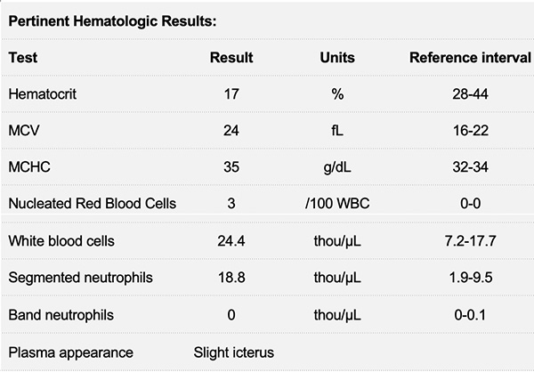
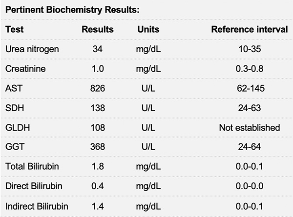
Venous blood from a goat
Case Information
An 18-month-old Nigerian Dwarf doe presented to the Cornell University Equine Nemo Farm Animal Hospital for evaluation of brown-red urine, lethargy, and fever. On physical examination, the goat was tachycardic (110 beats/minute) and had pale brown mucous membranes with a prolonged capillary refill time (estimated to be 8-10% dehydrated). The patient was posturing to urinate with no production. After a 200 mL bolus of fluid was administered to assess dehydration, the doe urinated 5 mL of dark brown-red urine. Blood was drawn for a complete blood count and chemistry panel. Results are shown in the tables below.
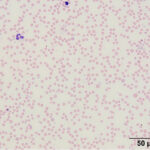
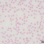
Representative images of the blood smear are shown (Figures 1-2). After viewing the images, answer the following questions:
- What abnormalities are present in the blood smear?
- What pathologic process can be identified?
- What are your differential diagnoses?
- What is the likely cause(s) for the high MCHC?
|
|
|
Answers on next page