
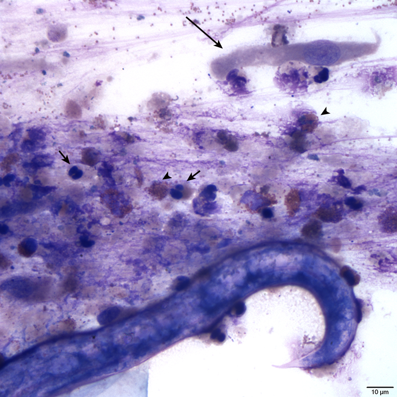
Figure 2. Tracheal wash sediment smear. High power image showing a columnar respiratory epithelial cell (long arrow), eosinophils (arrowhead), and poorly preserved neutrophils (short arrow). A portion of a lungworm larva can also be seen at the bottom of the image for size comparison (Wright’s stain, 50x).