
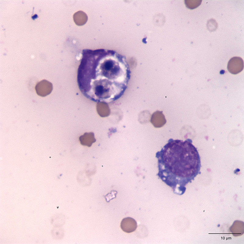
Figure 8: Percardial fluid, direct smear. Lymphoma cells, one of which is demonstrating cytophagic activity (top cell in image). (1000x, Wright’s stain)
Figure 8: Percardial fluid, direct smear. Lymphoma cells, one of which is demonstrating cytophagic activity (top cell in image). (1000x, Wright’s stain)
eClinpath helped 1.2 million visitors last year from 220 countries find important information on animal health. If you enjoy the site, please support our mission and consider a small gift to help us keep pace with its rapid growth. You can donate securely via PayPal or credit card. Thank you!
![]()