
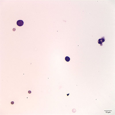
Figure 4: Pleural fluid, direct smear. A small and intermediate lymphocyte (cell on upper left) are evident, along with a neutrophil, which can be used for size comparison. (500x, Wright’s stain).
Figure 4: Pleural fluid, direct smear. A small and intermediate lymphocyte (cell on upper left) are evident, along with a neutrophil, which can be used for size comparison. (500x, Wright’s stain).
eClinpath helped 1.2 million visitors last year from 220 countries find important information on animal health. If you enjoy the site, please support our mission and consider a small gift to help us keep pace with its rapid growth. You can donate securely via PayPal or credit card. Thank you!
![]()