
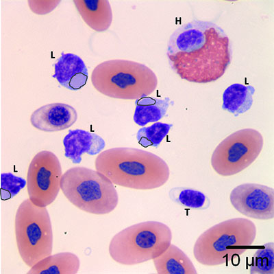
Figure 2b: The heterophil (H) has evidence of toxic change in the cytoplasm. Viral inclusions (black outline) are noted in some lymphocytes (L). A single thrombocyte (T) is also present (Wright’s stain, 1000x)
Figure 2b: The heterophil (H) has evidence of toxic change in the cytoplasm. Viral inclusions (black outline) are noted in some lymphocytes (L). A single thrombocyte (T) is also present (Wright’s stain, 1000x)
eClinpath helped 1.2 million visitors last year from 220 countries find important information on animal health. If you enjoy the site, please support our mission and consider a small gift to help us keep pace with its rapid growth. You can donate securely via PayPal or credit card. Thank you!
![]()