
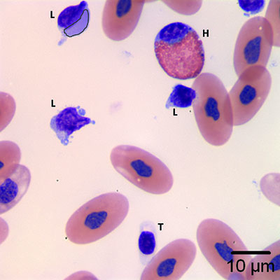
Figure 1b: This image contains numerous lymphocytes (L), with a heterophil (H) and a thrombocyte (T). An inclusion body is present in one of the lymphocytes (black outline). Wright’s stain, 1000x magnification.
Figure 1b: This image contains numerous lymphocytes (L), with a heterophil (H) and a thrombocyte (T). An inclusion body is present in one of the lymphocytes (black outline). Wright’s stain, 1000x magnification.
eClinpath helped 1.2 million visitors last year from 220 countries find important information on animal health. If you enjoy the site, please support our mission and consider a small gift to help us keep pace with its rapid growth. You can donate securely via PayPal or credit card. Thank you!
![]()