
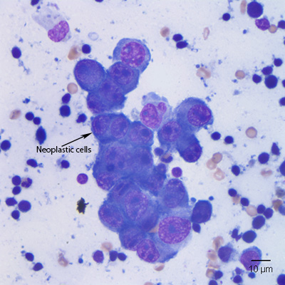
Figure 1: The neoplastic cells are cohesive, with a high nuclear to cytoplasmic ratio, 1-3 prominent large nucleoli, and a small amount of dark blue cytoplasm. No cytoplasmic pigment granules were identified (right mandibular lymph node, slide 1).
Figure 1: The neoplastic cells are cohesive, with a high nuclear to cytoplasmic ratio, 1-3 prominent large nucleoli, and a small amount of dark blue cytoplasm. No cytoplasmic pigment granules were identified (right mandibular lymph node, slide 1).
eClinpath helped 1.2 million visitors last year from 220 countries find important information on animal health. If you enjoy the site, please support our mission and consider a small gift to help us keep pace with its rapid growth. You can donate securely via PayPal or credit card. Thank you!
![]()