
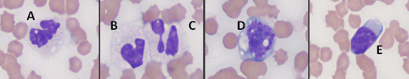
Figure 3: Image A is an “aged” segmented neutrophil with cytoplasmic vacuolation, mimicking toxic change. Image B is an “aged” segemented neutrophil with a swollen nucleus, that mimics a band neutrophil. Image C is a segmented neutrophil that is also demonstrating nuclear swelling (presumed secondary to imbibition of water). Both of the latter cells also demonstrate cytoplasmic vacuolation. Image D is a monocyte and image E is a lymphocyte.