
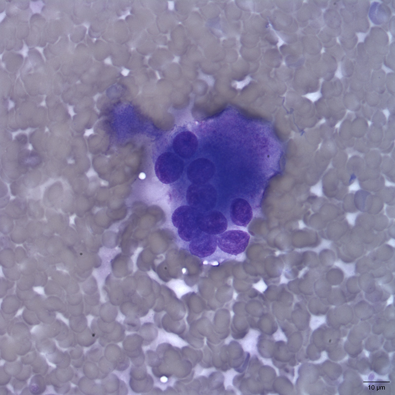
One of several mature osteoclasts seen in the aspirate of an aggressive bone lesion in a dog. Osteoclasts confirm the presence of osteolysis. The osteoclast in the image is interpreted as mature given its large size and multiple nuclei. The cytoplasm is also pink/magenta and grainy, which helps to distinguish it from a mutlinucleated macrophage. (Diff-Quik stain, 50x objective)