
Interpretation
Nasal cryptococcosis
Explanation
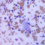
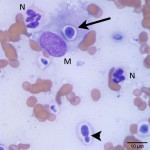
The most common causes of nasal disease resulting in facial deformity include neoplasia and fungal rhinitis, though severe chronic rhinitis is also possible (Question 1).1,2 The most common neoplastic processes affecting the nasal cavity in cats include lymphoma and epithelial tumors (carcinoma, adenocarcinoma and squamous cell carcinoma), though a variety of other tumors such as sarcomas, melanomas and mast cell tumors also occur in this location.3 Cryptococcosis is the most common cause of fungal rhinitis in cats, but other fungal agents such as Aspergillus spp. have been reported.2 The cytologic findings displayed in Figures 1b and 2b reveal many non-degenerate neutrophils (N) with fewer macrophages (M) and occasional lymphocytes (L), indicating a mixed neutrophilic, histiocytic (pyogranulomatous) inflammatory process (Question 2). Also present on the smear and occasionally within macrophages (arrow in Figure 2b) are many round to oval yeast organisms (Y) with a thick clear capsule. These organisms often display narrow-based budding (arrowhead in Figure 2b) and are compatible with Cryptococcus spp. (Question 3). Neoplastic cells were not identified.
|
|
|
Further information and follow up
Histopathology of the nasal mass confirmed the cytologic findings. The biopsy revealed infiltration of the nasal submucosa with large numbers of neutrophils, macrophages and fewer eosinophils, lymphocytes and plasma cells. Scattered throughout were numerous extracellular and intra-macrophagic yeast organisms surrounded by a thick clear capsule. A cryptococcal antigen titer was submitted and was positive (>2048). The patient was started on a course of antifungal mediation (fluconazole) and clinical improvement was seen within a week of starting treatment with a noticeable improvement in breathing effort.
Discussion
Cryptococcosis is caused by an encapsulated yeast organism acquired from the environment and is the most common systemic mycosis in cats. In most cats the disease is caused by either Cryptococcus neoformans or Cryptococcus gattii.2 In tissues, both of these organisms exist as round to oval yeast with the characteristic features of narrow based budding and a thick polysaccharide capsule (as seen in Figures 1b and 2b) and can only be distinguished by fungal culture. The capsule provides protection both from the environment, where it can survive for years, and from the host immune system. C. neoformans is distributed worldwide and has been found to reside in bird excreta and decaying plant matter. C. gattii was historically seen only in tropical and subtropical climates, but the distribution is now thought to include much of the world with an increased incidence of infections reported in humans and animals since 1999 in the Pacific Northwest of North America where the organisms have been isolated from tree bark, air and water samples.2,4
Although humans and other species are also at risk for cryptococcosis, cats appear to be more susceptible than many other species.2 Disease transmission between infected patients has not been reported and susceptible people and animals acquire the infection from shared environmental sources. Incubation times in animals can vary from 1 to 13 months or longer. Exposure with subclinical infection can also occur followed by recrudescence of the infection months or years later. This may have occurred in this patient since he had been kept indoors for the past five years, however, indoor cats can also become infected by aerosols from open windows or doors or exposure to indoor soil.4 The nasal cavity is the most commonly affected area in cats and is usually the primary site of infection. As a result, upper respiratory signs are frequent and generally chronic, including sneezing, nasal discharge, inspiratory dyspnea, stertor and the presence of mass-like lesions. Mass-like lesions can present as proliferative lesions protruding from the nares or as swellings over the bridge of the nose resulting in facial deformity, as seen in this patient. Spread through the cribriform plate or the auditory canal can result in central nervous system (CNS) signs or otitis media. Skin lesions and central nervous system (CNS) signs can also occur via hematogenous spread and ocular as well as systemic infections have been also been reported.2 Although clinical signs are similar for C. neoformans and C. gattii, infections with C. gattii are often more virulent and more likely to involve the CNS.4
Cytology is a rapid, inexpensive and sensitive diagnostic test for cryptococcosis. A diagnosis of cryptococcosis can often be made by identifying the organism in cytology samples from deep nasal swabs, nasal flushes and needle aspirates from lesions, but fungal culture is needed for speciation of the infection.4 Urine sediment should also be examined since some animals will have a renal infection and can shed organisms in the urine2 and organisms may be seen in cerebrospinal fluid (CSF) if CNS signs are present, however, collection of CSF is not always recommended due to the marked increase in CSF pressure in some patients.4 Histopathology can also be used for diagnosis and can help eliminate a neoplastic process in the nasal cavity. Special stains to highlight the characteristic polysaccharide capsule of the organism or immunohistochemical stains directed against the organism can be used for confirmation on formalin fixed tissue.5 A serum immunoassay for the cryptococcal polysaccharide capsular antigen is also available. This blood test is highly sensitive and specific for cryptococcosis and can help both confirm infection and provide a tool for monitoring the patient’s response to treatment. Titers usually increase initially with treatment due to dying organisms and should not be evaluated until 6-8 weeks after treatment is started. Treatment is continued until titers fall to 0.4
Treatment for cryptococcosis can have a good chance at success and cure, especially if started early in the disease process. Antifungal medications are the mainstay of treatment but surgical debulking of lesions is also indicated in some cases.2 Antifungal drugs used in feline cryptococcosis include the azoles: fluconazole, ketoconazole and itraconazole, with fluconazole recommended as the initial drug of choice given its low cost, tissue penetration characteristics and few side-effects.5 Resistance to fluconazole can be seen and some cats with severe disease or CNS disease (including many animals infected with C. gattii) will require other antifungal medications, such as amphotericin B and flucytosine.4 Treatment should be continued until clinical symptoms resolve, organisms are absent from infected tissues and serum cryptococcal antigen titers reach 0. Long term treatment, often 3-12 months and occasionally 2 years or more, with these drugs is required, which can be cost prohibitive for some owners. Although prognosis can be good to excellent depending on the distribution, extent and chronicity of the lesions, recurrences can still occur. Cryptococcal antigen titers should be monitored every 3-6 months in order to diagnose and treat relapses early.2,4
References
- Reed N. et al. Nasopharyngeal disease in cats: 1. Diagnostic Investigation. J Feline Med Surg 2012; 14(5): 306-315.
- Sykes J.E., Malik R. Cryptococcosis. In: Greene CE, ed. Infectious Diseases of the Dog and Cat. 4th ed. Philadelphia, PA: WB Saunders; 2011: 621-634.
- Withrow, S.J. Tumors of the Respiratory System. In: Withrow SJ, Vail DM, eds. Small Animal Clinical Oncology: 4th ed. St. Louis, MO: Saunders Elsevier; 2007: 535.
- Lester, S.J. et al. Cryptococcosis: update and emergence of Cryptococcus gattii. Vet Clin Path 2011; 40(1): 4-17.
- Trivedi S.R. et al. Feline Cryptococcosis: Impact of current research on clinical managment. J Feline Med Surg 2011; 13(3): 163-172.