
Interpretation:
Heatstroke resulting in acute renal, hepatocellular, muscle and gastrointestinal injury and disseminated intravascular coagulation
Explanation
Although the patient’s body temperature was not elevated at presentation, the provided history and physical exam findings indicate that she was suffering from severe heatstroke. The cooling performed by the owner prior to presentation likely returned her body temperature to normal but thermal damage had already begun. The number of nRBCs on the blood smear is striking (rubricytosis) and causes for this include a robust regenerative response, bone marrow injury, splenic contraction or altered splenic function and lead toxicosis. There is no evidence of a regenerative response in this case since only mild polychromasia is present on the smear and the patient is not anemic (she is actually polycythemic from dehydration and/or splenic contraction) and there was no history of lead exposure. Inappropriate rubricytosis is a very common finding in canine heatstroke and has been reported in 68% of dogs in one retrospective study.1 This finding has also been shown to be a sensitive and specific marker of death in dogs with heatstroke in another study, though it should not be relied upon as the sole prognostic indicator.2 The mechanism of the increase in nRBCs in this case may be direct bone marrow injury from hyperthermia but histologic correlates have not been able to confirm this mechanism.3 Other possibilities include splenic contraction in the early phases of the condition due to poor visceral perfusion or cytokine mediated release of nRBCs from the systemic inflammatory response.4 The WBC count obtained from the automated analyzer needs to be corrected for nRBCs since all nucleated cells are included in the WBC count and the presence of nRBCs will falsely increase the WBC count. The following calculation is used to correct the WBC count:
Corrected WBC count = Automated WBC count x 100/(100+nRBC)
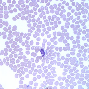
The cells identified by arrowheads in Figure 1b are apoptotic leukocytes, likely neutrophils. Large proportions of the leukocytes on the smear were apoptotic, karyolitic or had botryoid nuclei (having nuclear segments clustered similar to a bunch of grapes – see Figure 2). These changes are also a result of the heatstroke and have been reported in the literature as a consequence of hyperthermia in dogs.4,5 Assessment of the chemistry panel indicates that the organs affected include the kidneys (increases in urea, creatinine), the liver (increases in ALT, AST) and muscle (increases in AST, CK). The alterations in plasma proteins with decreases in both albumin and globulin (day 2) along with the clinical signs of vomiting and diarrhea with hematochezia also indicate gastrointestinal injury. The platelet count was within the reference limits on day 1 but by day 2 had dropped to below 30 thousand/uL. The concurrent increase in D-Dimer, APTT and PT and the decrease in fibrinogen and antithrombin indicate that the mechanism of the thrombocytopenia is consumption from disseminated intravascular coagulation (DIC). Other mechanisms such as heat-induced vasculitis, hyperthermia induced platelet aggregation and destruction are also possible.
Discussion
Heatstroke is relatively common in dogs, especially in the warmer summer months and can result from exposure to a hot environment, such as this case, but also from strenuous physical exertion. It is characterized by high core temperatures of >41 C (105.8 F) in dogs and often associated with systemic inflammatory response syndrome and leads to multiple organ dysfunction and death.1 During hyperthermic states and exertion, blood shifts from the mesenteric circulation to the muscles and skin in order to meet oxygen demands and dissipate heat resulting in intestinal ischemia, hypoxia and hyperpermeability explaining the gastrointestinal signs in this patient and in others.3 The many other lesions of heatstroke relate to direct thermal insult to tissues, as well as secondary effects of dehydration, shock and poor organ perfusion.1 Thermal endothelial injury activates the coagulation and complement cascades and often results in DIC and a systemic inflammatory response syndrome. Acute renal injury occurs due to both prerenal and renal mechanisms, including dehydration, direct thermal injury to renal tubules resulting in tubular necrosis as well as hypoxia, endotoxemia and release of cytokines and vasoactive substances. Microthrombi can also form resulting in thrombosis and ischemia of the kidneys and of other organs such as the liver and muscle.1 Muscle injury explains the marked increase in CK in the initial 24 hours of treatment of this patient and the resultant rhabdomyolysis can also contribute to the renal injury by release of myoglobin and subsequent myoglobinuric renal injury.1 Cardiac muscle effects can also result in arrhythmias from both direct thermal injury and from the secondary effects of hypoperfusion, lactic acidosis, electrolyte imbalances and thrombosis resulting in myocardial ischemia. 1,3
In a retrospective study of 54 dogs suffering from heat stroke 78% of cases occurred in June, July or August.1 Although heat stroke is characterized by two major findings: 1) an elevated core temperature and 2) central nervous system abnormalities and dysfunction,3 variability in the body temperature on admission is often seen6 and the canine brain has been shown to be more resistant to thermal injury than the human brain7 making these criteria less reliable in detection of heatstroke in dogs. Some animals present either normothermic (as was seen in this case) or hypothermic following a heatstroke event,2 possibly due to delay in admission to hospital and prior body cooling by owners or referring veterinarians. As such, a high body temperature should not be the only criteria used to identify dogs with heatstroke. In fact, no association between the body temperature on admission and survival was found in one retrospective study.1 Other historical and laboratory data and clinical findings such as exposure to high environmental temperatures and/or strenuous activity, the presence of inappropriate rubricytosis or apoptotic neutrophils with or without botryoid nuclei and a neurologically abnormal dog should alert practitioners to the possibility of heatstroke even in the absence of hyperthermia.
Overall mortality rate in heat stroke in dogs has been shown to be about 50% and early diagnosis and intervention is critical for the successful treatment of this condition.1 Negative prognostic indicators include delayed admission to hospital (beyond 90 minutes), hypoglycemia, prolonged PT and APTT at admission, serum creatinine >1.5 mg/dL after 24 hours, seizures, obesity, 1 and the relative and absolute number of nRBCs in peripheral blood.2
Aggressive treatment with intravenous fluids to restore circulatory volume and whole body cooling to inhibit fibrinolysis are important, but treatment for DIC is critical including administration of fresh frozen plasma. Antibiotics are also indicated to combat endotoxemia from gastrointestinal injury and other common medications include the use of mannitol, H2-receptor blockers, furosemide, glucose, dexamethasone, dopamine and diazepam. 1
Further information and follow up
The patient was treated aggressively with intravenous fluids, fresh frozen plasma, antibiotics, antiemetics and analgesics. Monitoring of renal analytes over the course of 3 days revealed steady increases in serum creatinine from 1.9 mg/dL at admission to 9.1 mg/dL by day 3 (Ref 0.6-1.4). Given the poor prognosis associated with worsening of the renal parameters the decision was made to euthanize the patient.
This case illustrates the devastating effects of heatstroke in dogs. Given the fact that history in cases of heatstroke can be lacking, recognition of the typical hematologic abnormalities seen in this condition can aid in early intervention and improve survival times in these patients.
References
- Bruchim Y, Klement E, Saragusty J, et al. Heat stroke in dogs: A retrospective study of 54 cases (1999-2004) and analysis of risk factors for death. J Vet Intern Med 2006;20:38-46.
- Aroch I, Segev G, Loeb E, et al. Peripheral nucleated red blood cells as a prognostic indicator in heatstroke in dogs. J Vet Intern Med 2009;23:544-551.
- Bruchim Y, Loeb E, Saragusty J, et al. Pathological findings in dogs with fatal heatstroke. J Comp Pathol 2009;140:97-104.
- Mastrorilli C, Welles EG, Hux B, et al. Botryoid nuclei in the peripheral blood of a dog with heatstroke. Vet Clin Pathol 2013;42:145-149.
- Wilcox A, Russell KE. Hematologic changes associated with Adderall toxicity in a dog. Vet Clin Pathol 2008;37:184-189.
- Drobatz KJ, Macintire DK. Heat-induced illness in dogs: 42 cases (1976-1993). Journal of the American Veterinary Medical Association 1996;209:1894-1899.
- Oglesbee MJ, Alldinger S, Vasconcelos D, et al. Intrinsic thermal resistance of the canine brain. Neuroscience 2002;113:55-64.
Authored by: Dr. H. Priest