
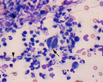
Figure 2b: This image reveals similar inflammatory cells, but several non-staining fungal hyphae (arrowheads) are evident in the smear, one of which appears to be phagocytized within a macrophage (arrow). The macrophage is also binucleate (Wright’s stain).