
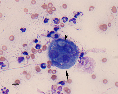
Many non-degenerate neutrophils are present in the smear, along with background erythrocytes. There is a single multinucleated macrophage (arrow), which appears to contain a phagocytized fungus with a bulbous end (arrowhead) (Wright’s stain).
Many non-degenerate neutrophils are present in the smear, along with background erythrocytes. There is a single multinucleated macrophage (arrow), which appears to contain a phagocytized fungus with a bulbous end (arrowhead) (Wright’s stain).
eClinpath helped 1.2 million visitors last year from 220 countries find important information on animal health. If you enjoy the site, please support our mission and consider a small gift to help us keep pace with its rapid growth. You can donate securely via PayPal or credit card. Thank you!
![]()