
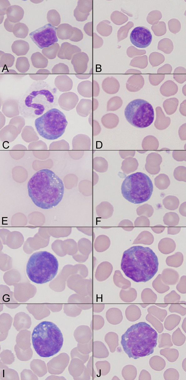
Reactive lymphocytes (all taken at the same magnification) in a older dog with an atrial hemangiosarcoma.
A: Normal small, mature lymphocyte. B: Small “plasmacytoid” lymphocyte: this cell has increased amounts of darker blue cytoplasm than normal. C: Another variant of a “plasmacytoid” lymphocyte. The adjacent neutrophil indicates the lymphocyte is small. D: Another variant of a “plasmacytoid” lymphocyte with more abundant cytoplasm and a perinuclear clear zone. E: A large reactive lymphocyte. This cell has cytoplasmic vacuoles, medium blue cytoplasm, but a coarse chromatin with no nucleoli. F: A larger “plasmacytoid” lymphocyte. G: Large reactive lymphocyte with a pleomorphic nucleus and smooth dark blue cytoplasm. H: Large reactive lymphocyte with “bi-lobed” nucleus. I: Large reactive lymphocyte with pleomorphic nucleus, deep blue cytoplasm and cytoplasmic vacuoles. J: Another variant of a large reactive lymphocyte with a pleomorphic nucleus.