
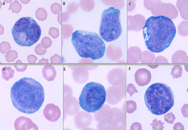
This image illustrates the difficulty in distinguishing between reactive lymphocytes and neoplastic hematopoietic cells. These six images are snapshots of “big blue” cells from dogs. Three were reactive (A, C, E) – these cells have relatively clumped chromatin (heterochromatin) and mostly smooth blue cytoplasm. The cell in E is actually a plasmacytoid lymphocyte (resembles a plasma cell) and is not as large as the other cells in the image. The other three cells (B, D and F) are from dogs with leukemia. B are cells from a dog with acute myeloid leukemia (the cells were monoblasts on immunuphenotyping and cytochemical staining) – one has a distinct nucleolus and the two cells look more alike than different. D is from a dog with acute T lymphoid leukemia (based on positive reactions for CD3 and CD5 on flow cytometric immunophenotyping and negative reactions for cytochemical stains). F is a cell from a dog with lymphoma. This has relatively clumped chromatin and, at first glance, appears reactive, however more cells of this kind were seen in blood and resembled those in the dog’s lymph node aspirate. This suggests this was a neoplastic and not reactive lymphocyte.