
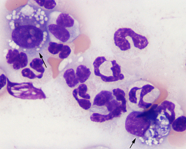
Two histiocytes (arrows) are seen at the feathered edge of a blood smear in a dog with inflammation due to pancreatitis. The cells resemble macrophages and not monocytes because they have round to oval eccentric nuclei with a moderate amount of vacuolated cytoplasm and are demonstrating phagocytic activity (particularly the cell on the lower right) (Wright’s stain, 1000x magnification).