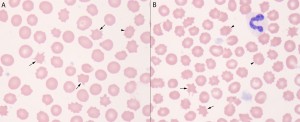
Acanthocytes are irregularly spiculated cells (spicules are irregular in size, shape and distribution around the RBC membrane), whereas echinocytes are regularly spiculated cells. In this compilation of images from a blood smear of a dog with disseminated intravascular coagulation, acanthocytes are seen (arrows, A and B). Some cells also have more regular membrane projections (arrowheads, A and B). Although these could be variants of acanthocytes, these are echinocytes. This demonstrates that these two RBC shapes can be difficult to distinguish from each other. Of these RBC shape changes, acanthocytes are of more pathologic relevance. In this case, they should raise the index of suspicion of DIC (because the dog is concurrently thrombocytopenic (with large platelets) and other fragments are present (keratocytes). A neutrophil is also present in image B (Wright’s stain, 1000x magnification).

