
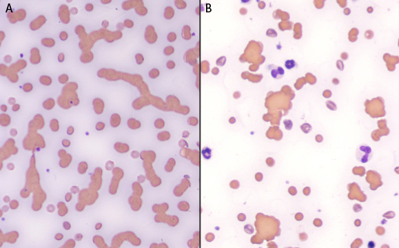
A: This image demonstrates rouleaux formation (RBC forming stacks of variable length) in a cat with a monoclonal gammopathy due to multiple myeloma. The blue background is also a reflection of the high protein in the sample 911.5 g/dL).
B: This image demonstrates three-dimensional clumps of RBC, compatible with agglutination, in a blood smear of a dog with immune-mediated hemolytic anemia. This clumping did not dissipate with saline dilution, whereas the rouleaux formation in the cat did (not shown).