
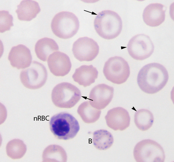
In this blood smear from a dog with chronic lead poisoning, the lead has resulted in a “relative” iron deficiency anemia, with several hypochromic RBC (which are also target cells, arrowhead, because the cells are thinner than normal). Lead inhibits the incorporation of iron into heme, so the iron accumulates in mitochondria resulting in the formation of RBC containing iron inclusions or siderocytes (arrows). A high number of nucleated RBC (nRBC), disproportionate to the degree of polychromasia and basophilic stippling (lead inhibits the enzyme 5′ nucleotidase which degrades ribosomes) results in basophilic stippling in mature, polychromatophilic RBC (B) and nRBC (Wright’s stain, 1000x magnification).