
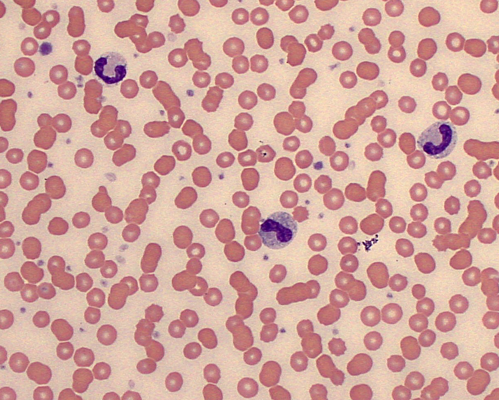
The dog had a normal leukocyte and neutrophil count but more band neutrophils than segmented neutrophils (degenerative left shift) with marked toxic change as illustrated in this image (marked cytoplasmic basophilia, cytoplasmic vacuolation and Dohle bodies). The dog had a metritis. The cells going from left to right are a segmented neutrophil, a band neutrophil to metamyelocyte, and a band neutrophil. There is background rouleaux formation, likely due to increased fibrinogen (inflammation) or immunoglobulins (polyclonal; antigenic stimulation).