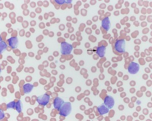
In this venous blood smear from a dog, there are many large mononuclear cells with fine chromatin and indistinct nucleoli. The cells have mostly round nuclei (some nuclei are pleomorphic) with small amounts of light blue cytoplasm. A few cells have purple cytoplasmic granules (arrow), which are more typical of myeloid blasts (particularly mono blasts) versus blasts of lymphoid origin cells. The results indicate an acute leukemia (comprised of immature cells). Flow cytometric analysis and cytochemical staining confirmed an acute monoblastic leukemia (the neoplastic cells displayed markers of monoblasts including CD11b, CD11c and alkaline phosphatase). The dog was concurrently pancytopenic (neutropenia, non-regenerative anemia, thrombocytopenia). Bone marrow aspiration revealed >80% blasts that were similar to those seen in blood (Wright’s stain, 50x magnification).

