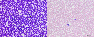
Overstaining a smear can make it difficult to identify cells, such as polychromatophils and monocytes from neutrophils. Cellular features such as tocic change in neutrophilsa re difficult to discern. Left image: A smear of canine blood that was overstained (likely in the blue dye) with a rapid dye, such as Diff-quik. Erythrocytes are quite dark. A band neutrophil (9 o’clock), segmented neutrophil and lymphocyte (arrow) can be identified. The neutrophils have a falsely “grainy” cytoplasm. Right image: The same smear was stained with a modified Wright’s stain showing how much easier it is to identify cells (band neutrophil to the left, neutrophil to the right) as well as their cytoplasmic features.

