
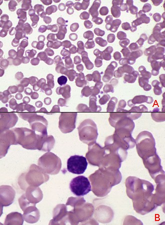
These pictures show the appearance of the portion of the blood smear that is too thick to be used for identifying and assessing blood cells. The thick area of a smear dries too slowly for leukocytes to spread out. The white cells here are shrunken and appear much smaller than in the quickly dried areas. There is little or no definition of features; cytoplasm and nucleus cannot be distinguished.
The white cells shown in the bottom panel might be either lymphocytes or monocytes but they are too shrunken to display features that would allow identification. The red cells are stacked in long rouleaux in this area. Potentially important morphologic abnormalities are not visible in slowly dried cells in the thick area of the smear.